Product Description
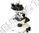
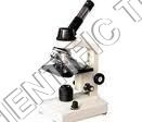
We have emerged in this domain as one of the prime manufacturers, suppliers and exporters of Trinocular Microscope. These products are used for magnifying various types of specimens in laboratories and medical institutes. Equipped with three ports, these products facilitate excellent viewing and detection experience. A camera can be attached to the third port in order to take images of the specimen. Packed in damage free packaging material, Trinocular Microscope is delivered to the clients in a secure manner.
Key Features:
- High quality Trinocular Microscope for School, Colleges and Research Institutes.
- Provided with a Trinocular head binocular observation inclined at 45degree,
- with interpupillary distance 55 mm to 75 mm & diopter adjustments and a straight tube for photomicrography.
- Rectangular 115 x 125 mm with co-axial mechanical stage.
- Sub stage movable by rack and pinion Abbe Condenser of 1.25 N.A. with iris diaphragm and filter
- Box type base with 6v-20w, variable halogen illumination.
- Achromatic objectives:4x, 10x, 40x, 100x(SL).
- Eyepieces:10x(pair) WF
With a double-layer mechanical stage featuring a graduated, smooth XY movement of 75mm x 50mm, users enjoy effortless specimen navigation for precise observation and measurements. The stage's ergonomic controls are suitable for repeated use in academic and research laboratories.
Advanced Optical and Digital ImagingEquipped with achromatic objectives (4x, 10x, 40x spring, 100x oil spring) and wide field eyepieces, this trinocular microscope delivers crisp images at 40x to 1000x magnifications. The standard C-mount and USB interfaces provide seamless integration with CMOS cameras for enhanced digital documentation.
Stable, Durable ConstructionThe robust, corrosion-resistant metal body and anti-vibration feet ensure long-term stability and reliability. Designed for intensive laboratory environments, the microscope's ergonomic frame supports comfortable handling and longevity, even under demanding conditions.
FAQ's of Trinocular Microscope:
Q: How do I use the graduated stage movement for precise specimen positioning?
A: The graduated, smooth XY stage movement (75mm x 50mm) allows for exact positioning and navigation of specimens, facilitating detailed observation and imaging. You operate the stage using ergonomic controls, which provide incremental movements ideal for repetitive laboratory tasks.
Q: What features enhance digital imaging with this trinocular microscope?
A: The microscope offers a standard C-mount camera port, compatibility with CMOS sensors, and USB 2.0/3.0 interfaces for digital models. These features enable high-resolution imaging up to 5MP, with still image formats in JPEG/BMP and video capture at up to 1920x1080 resolution.
Q: When should the recommended cover glass thickness be used for optimal results?
A: For the best image quality, use a cover glass thickness of 0.17mm, as specified for this microscope model. This dimension matches the microscope's optical parameters and minimizes distortion or loss of resolution during specimen observation.
Q: Where can this trinocular microscope be utilized effectively?
A: This microscope is designed for laboratory use and excels in settings such as research institutes, universities, medical labs, and industrial inspection sites across India. Its durability and advanced imaging capabilities make it suitable for both educational and professional environments.
Q: What is the process for capturing and saving images or videos during specimen analysis?
A: To capture images or videos, connect a compatible digital camera using the C-mount and USB interface. Still images can be saved in JPEG or BMP formats, while videos are recorded in AVI, allowing for documentation and sharing of your observations.
Q: How does the anti-vibration feature benefit laboratory work?
A: The anti-vibration feet fitted to the microscope stabilize the instrument during use, reducing image blur and enhancing precision when focusing or capturing digital images. This feature is particularly beneficial during high-magnification studies and long observation periods.