Product Description
Our modern infrastructure unit and technical know how enables us to manufacture, supply and export Fluorescent Microscope. These products are widely used for the absorption and reflection of properties in organic and inorganic substances. Equipped with phosphorescence and fluorescence, these products provide correct and quick results. Known for their effective functioning and user friendly controls, these products have earned the trust of the clients in the international arena. Fluorescent Microscope is available with us in different specifications as per the needs of the clients.
Specifications:
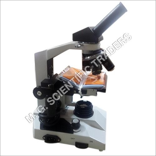
Infinity Optical System (IOS)
Siedentopf observation head inclined at 30degree rotatable at 360degree. IPD 55-75 mm,
with Dioptric adjustment
EYEPIECE
Wide field eyepiece 10X (Paired) FOV 22mm
OBJECTIVES INFINITY CORRECTED
Fluorescence objective 4X
Fluorescence objective 40X (SL)
Fluorescence objective 10X
Fluorescence objective 100X (SL) Oil
MECHANICAL BODY
Co-axial focusing system with large knobs, PRE-FOCUSING LEVER & TENSION.
ABBE condenser NA 1.25
ADJUSTMENT RING.
Reverse Angle quadruple nosepiece on ball bearing.
Mechanical stage is 185mmX142mm.
The Mechanical stage has low positioned co-axial controls on ball bearing guide ways.
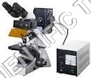
LIGHT SOURCE : 12V, 20W halogen illumination
Epi - Fluorescent illumination :
Blue Excitation Light Filter System (400-490nm) Standard
Green Excitation Light Filter System (510-550nm) Standard
100 WHBO Ultra Hi-Voltage Spherical Mercury Lamp, with power supply.
Transmitted 6V/30W Tungsten Halogen bulb.
Protection Barrier to Resist the Ultra Violet Light with Power Supply & four Fluorescence Free objectives: FL 4X, FL 10X, FL 40X (S), FL 100X (S, Oil).
The Epi-Fluorescent attachment can be flexibly equipped with ordinary microscope to be an Epi-Fluorescent microscope. The Fluorescent microscope is used to detect bacteria and antigens, early diagnosis of cancer, venereal diseases, immune less diagnosis, examination of botanical materials and the study of viruses etc.
Superior Imaging PerformanceEquipped with plan achromatic objectives and a high-sensitivity CCD or CMOS sensor, the microscope delivers sharp, high-resolution images up to 1920 x 1080 pixels. Multiple filter sets enable selective fluorescence excitation, providing clear visualization of specimens. The trinocular head enhances both observation and documentation with dedicated photo ports.
User-Friendly and Ergonomic DesignThe microscope boasts a double-layer mechanical stage, inclined eyepiece tube, and 360 rotation for comfortable extended use. Coarse and fine focus options with tension adjustment allow precise focusing, while wide field eyepieces and adjustable interpupillary distance accommodate different users. Anti-fungal coatings and stainless steel material ensure long-term integrity.
Flexible Connectivity and OutputUsers can easily capture and export still images and full HD video via USB, HDMI, or AV interfaces. The system supports common image formats, including JPG, BMP, and TIFF. Real-time video streaming (up to 60 fps) and high-resolution still capture make documentation and analysis efficient for laboratory workflows.
FAQ's of Fluorescent Microscope:
Q: How is stage movement controlled on the fluorescent microscope and why is it beneficial for specimen observation?
A: Stage movement is managed via a double-layer mechanical stage, offering X-axis movement of 75 mm and Y-axis movement of 50 mm. This precise control ensures accurate specimen positioning, facilitates scanning across slides, and allows efficient observation of targeted areas-crucial for detailed fluorescence studies.
Q: What imaging options are available on this microscope and how are images exported for analysis?
A: The microscope integrates high-sensitivity CCD or CMOS sensors with resolutions up to 1920 x 1080 pixels (Full HD). Images and videos can be exported via USB, HDMI, or AV outputs, in formats like JPG, BMP, and TIFF, enabling easy documentation, sharing, and analysis.
Q: When should laboratory users select different filter blocks for fluorescence studies?
A: Different filter blocks-B, G, U, V-are chosen based on the fluorescent dyes or markers used in the specimen. Each set selectively excites specific wavelengths, ensuring optimal visualization. Users should select corresponding filter blocks aligned with the emission spectra of their labels for best results.
Q: Where is this microscope manufactured and distributed, and who can purchase it?
A: This microscope is manufactured and distributed in India. It is available through various channels, including dealers, exporters, manufacturers, suppliers, retailers, traders, and wholesalers. Laboratories and research institutes can procure it conveniently across the country.
Q: What is the process for adjusting focus and illumination on the microscope during use?
A: Focus is managed through a coaxial system with both coarse (30 mm range) and fine (0.2 mm range) adjustment, allowing precise specimen clarity. Illumination uses a high-intensity LED or optional mercury lamp for reflected and transmitted light, both controlled to provide optimal viewing for fluorescence observation.
Q: How does the microscope's ergonomic design contribute to user comfort during laboratory work?
A: The microscope features an inclined eyepiece tube set at 30, 360 rotatable view head, and wide interpupillary adjustment (48-75 mm). These ergonomic elements reduce fatigue during extended sessions, ensure comfortable observation angles, and accommodate multiple users efficiently.