Product Description
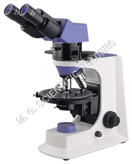
We have acquired a rich domain expertise to undertake and successfully manufacture, supply and export Polarizing microscope. These products are used in different laboratories and medical colleges for viewing objects under polarized light. Owing to their high strength and low maintenance, these products are very popular among our clients in the international market. Polarizing microscope can be availed from us in different models and specifications as per the requirements of the clients.
Specifications:
Eyepiece: WF 10X-20mm
Reticule Eyepiece: WF 10X-20 mm (Cross Reticule 0.1mm)
Objective: Infinity LONG WORKING DISTANCE PLAN Objective , 4X/0.1,10X/0.25, 20X/0.4 (S) and 40X/0.65 (S)
Head: Seidentopf Binocular / Trinocular Head Inclined 30, Rotatable 360, Interpupillary Distance: 50-75mm
Nosepiece : Quadruple
Revolving Mechanical Stage: Rotatable 360, Center Adjustable, Division 1, Vernier division 6
Condenser: Abbe N.A. 1.25 with Iris Diaphragm
Focus: Coaxial Coarse and Fine Focusing Knobs
Illumination: LED 3.3W/3V, ISO - CE
Filter: Amber, green and Neutral
Polarizing system: Polarizing Analyzer, Bertrand Lens, Slip-Red,1/4Slip,Quartz Wedge.
Digital Eyepiece camera 3.0 MP, USB 2.0
Sensor Parameter: 1/2" 3.0MP CMOS
Resolution: 2048*1536
USB port: USB 2.0
Frame Rate: 7.5f/s@ 2048*1536
Preview Model: 2048*1536,1280,1024 ,102,768
Pixel Size: 3.18m *3.18m
Sensitivity: 0.9v/lux sec
S/N Ratio: 40 dB
Dynamics Range : 60dB
White Balance: AUTO
The Length of USB: 1.5 meter
O/P compatibility: Windows--XP2, 7 and 8
Power supply USB port
Tube diameter: 23.2 mm or 30.00mm
Connection: Ocular tube or C-mount
Uniform focusing: YES
Optical magnification: 10X
Rate of field: 1/3
Matching lens: YES
Function of software:
Image file management: set up, open up, save, lead in image file
Image capture: Switch, setting, shoot, measure, full screen view.
Image edit: Picture turns, zoom, cut, modify, email and print.
Resolution of display: 2048*1536
Resolution of picture: 2048*1536
Resolution of video: 2048*1536
Weight of device: 0.10
Advanced Rotatable Polarizer and AnalyzerThe built-in polarizer and 360 rotatable analyzer are ideal for examining birefringent samples under varying polarized light orientations. This allows precise identification and study of optical properties in minerals and crystals, enhancing analytical capability for geological and material science laboratories.
Precision Stage and Optics for Reliable ResultsThe circular stage, graduated to 0.1 with vernier scale, offers exceptional accuracy when analyzing specimen orientation. Achromatic objectives and wide field eyepieces ensure sharp, clear visuals, while the coaxial coarse and fine focus delivers precise control and reproducible measurements.
Versatile Observation and Documentation OptionsA trinocular drawtube facilitates both comfortable visual examination and easy digital image or video documentation via an accessory camera mount. This flexibility supports teaching, research, and reporting requirements in a diverse laboratory environment.
FAQ's of POLARIZING MICROSCOPE:
Q: How does a polarizing microscope help in analyzing geological specimens?
A: A polarizing microscope utilizes polarized light to reveal optical properties specific to birefringent materials like minerals and crystals. This method enables geologists and material scientists to examine internal structures, identify compositions, and analyze physical properties that are otherwise invisible under standard brightfield illumination.
Q: What procedures should be followed to use the rotatable analyzer effectively?
A: To use the rotatable analyzer, place your specimen on the circular stage and rotate the analyzer through 360 while observing changes in brightness and coloration. This helps differentiate minerals by their optical behavior and is essential for methods such as conoscopic and orthoscopic observation.
Q: When is the Bertrand lens used during observation?
A: The built-in Bertrand lens is engaged for conoscopic observation, typically when analyzing interference patterns or optic axes in crystals. Swing the lens into the light path to view interference figures, and out for standard orthoscopic (direct) viewing of the sample structure.
Q: Where can digital images or videos be captured from the microscope?
A: The trinocular drawtube includes a mechanical accessory port that supports optional camera mounts. This allows the user to capture high-resolution still images or videos of microscopic specimens for further analysis or documentation.
Q: What are the key benefits of the graduated rotatable stage for laboratory users?
A: The circular stage, graduated to 0.1, lets users measure and track precise specimen orientation, which is crucial for comparative studies or repetitive analyses. The vernier scale ensures accurate readings, supporting quantitative research and teaching applications.
Q: How do you maintain the microscope for long-term usage?
A: Regularly clean the optics with lens paper, keep the instrument covered with the provided dust cover when not in use, and use only the recommended immersion oil. Check and replace spare parts like halogen lamps and fuses as listed in the user manual to maintain optimal performance.